Connection of the spinal column with the skull. Controls of the vertebrae Connection of the skull to the 1st cervical vertebra of the condyle in the occipital bone with the upper joint fossa of Atlanta form
Connection of the spinal column with the skull. Control compounds Connection of the skull with 1 cervical vertebra
Connections of the touch in the occipital bone with the upper articular fossa of Atlanta form a combined ellipse Atlanto -aligned joint (Articulatio Atlantoocpipitalis). In the joint, movements around the sagittal axis are possible – tilting the head to the sides and around the front axis – flexion and extension. The compound of Atlanta and the axial vertebra forms 3 joints: pair combined flat Lateral Atlanto -axis joint (Articulatio Atlantoaxial Lateralis), located between the lower articular surfaces of Atlanta and the upper joint surfaces of the axial vertebra; Non -pair cylindrical Middle Atlanto -axis joint (articulatio atlantoaxialis medialis), Between the tooth of the axial vertebra and the joint hole of Atlanta. The joints are strengthened by strong ligaments. Between the front and rear arcs of Atlanta and the edge of the large occipital opening are stretched Front and rear Atlantoisy membranes (Mambranae Atlantoocipitales Anterior et Posterior) (Fig. 36). Between the side masses of Atlanta throwsThe transverse ligament of Atlanta (Lig. Trasversum Atlantis). Fibrous passes through the upper free edge of the transverse ligament
Rice. 36. The connection of the cervical vertebrae with each other and with the skull: a – the cervical spine, view on the right side: 1 – intellectual ligament; 2 – yellow ligaments; 3 – exit ligament; 4 – the rear Atlantoisy membrane; 5 – anterior Atlantostya membrane; 6 – anterior longitudinal ligament;
b – the upper part of the spinal canal, view of the back. The arcs are removed
and spinous processes: 1 – lateral Atlantosa joint; 2 – Atlantoisy joint; 3 – occipital bone; 4 – cover membrane; 5 – rear longitudinal ligament; B – compared with the previous pattern, the cover membrane is deleted: 1 – the transverse ligament of Atlanta; 2 – porovable ligaments; 3 – cross -shaped ligament of Atlanta; g – compared with the previous pattern, the cross -shaped ligament of Atlanta is deleted:
1 – ligament of the top of the tooth; 2 – patch ligament; 3 – Atlantoisy joint; 4 – lateral Atlanto -axis joint;
D – Middle Atlanto -axis, View from above: 1 – transverse ligament of Atlanta;
2 – Prochite ligament
litter to the anterior semicircle of a large occipital hole. From the lower edge of the same ligament down, to the body of the axial vertebra, there is a fibrous bundle. The upper and lower bundles of the fibers, together with the transverse ligament, form The cross -shaped bunch of Atlanta (Lig. Cruciforme Atlantis). Two from the upper part of the side surfaces of the dent Poemic ligaments (Ligg. Alaria), heading to the muscles of the occipital bone.
The spinal column in general
The spinal column (columna vertebralis) It consists of 24 true vertebrae, sacrum, coccyx, intervertebral discs, articular and ligaments. The functional value of the spine is enormous. It is a container for the spinal cord lying in the spinal canal (Canalis Vertebralis); serves as a support of the body, participates in the formation of the thoracic and abdominal walls.
There are intervertebral openings between the above and the underlying vertebrae (Forr. InterteverTebralia), Where spinal nodes lie, vessels and nerves pass. The intervertebral holes are formed by the lower clipping of the overlying vertebra and the upper cut of the underlying.
The human spine has bends in the sagittal plane (see Fig. 18.1). In the cervical and lumbar sections, the spine forms bends directed by the bulge of the anterior lordosis (lordosis), And in the thoracic and sacral departments – bends directed back – kyphosis (Kyphosis). The bends of the spinal column give him spring properties. Bends are formed in the postnatal period. At the 3rd month of life, the child begins to raise his head, a cervical lordosis appears. When the child begins to sit, chest kyphosis forms (6 months). When moving to a vertical position, lumbar lordosis (8-9 months) occurs. The final formation of bends ends by the age of 18. Side bends of the spine in the front plane – Scoliosis – are pathological curvatures. In old age, the spine loses physiological bends, as a result of losing elasticity, a large breast bend, the so -called senile hump, forms. In addition, the length of the spine can decrease by 6-7 cm. Movements in the spinal column are possible around 3 axes: frontal – flexion and extension, sagittal – tilt to the right and left, vertical – rotational movements.
The connections of the vertebrae in the spinal column should, in addition to high mechanical strength, provide the spine with flexibility and mobility. These problems are solved due to the special method of joints of the articular surfaces of the vertebrae, as well as the location of the ligaments that strengthen these compounds. The intervertebral discs located between the vertebrae of the vertebrae (Discus intervertebralis), consisting of a fibrous ring (Annulus fibrosus) (Fig. 12), surrounding the so -called jet nucleus (nucleus pulposus) (Fig. 12), increase the stability of the spine to vertical loads and amorsate mutual mixtures Callors.
The combination of articular processes of the vertebrae is called artot -resistant connection (articulatio zygapophysialis) (Fig. 12). The joint is flat, formed by the articular surfaces of the upper articular processes of one vertebra and articular surfaces of the lower articular processes of the other – the overlying – vertebra. The joint capsule is attached along the edge of the articular surfaces. Each arched joint allows insignificant sliding movements, but the addition of these movements along the entire length of the spine gives it significant flexibility.
Rice. 12.
Arched compound (intervertebral connection between II and III lumbar vertebrae)
1 – upper joint process of the III lumbar vertebra;
2 – lower articular process of the II lumbar vertebra;
3 – arched joint;
4 – yellow ligament;
5 – transverse process of the III lumbar vertebra;
6 – rear longitudinal ligament;
7 – a jacket nucleus;
8 – fibrous ring;
9 – front longitudinal ligament
The arcs of adjacent vertebrae are interconnected by a yellow ligament (Lig. Flavum) (Fig. 12), transverse processes are connected by inter -divided ligaments, the gaps between the spinous processes are occupied by the intellectual ligaments that form a prejudicious ligament passing above the tops of the spinous processes. In addition, on the front surface of all vertebrae from the sacrum to the occipital, the anterior longitudinal ligament passes (Lig. Longitudinale Anterius) (Fig. 12). The rear surfaces of the vertebral bodies (from sacrum to II cervical) are connected by the posterior longitudinal ligament (Lig. Longitudinale Posterius) (Fig. 12). The front and rear longitudinal ligaments collect the vertebral column into one.
Rice. thirteen.
Connections between the occipital bone and I-II cervical vertebrae
1 – porovable ligaments;
2 – occipital bone;
3 – occipital taps;
4 – Atlantoisy joint;
5 – transverse process of Atlanta;
6 – lateral mass of Atlanta;
7 – cross -shaped ligament of Atlanta;
8 – lateral Atlanto -axis joint;
9 – body of the II cervical vertebra
A special type of connection is present at the junction of the upper vertebrae with the base of the skull.
The joint of the lateral masses of the I cervical vertebra (Atlanta) with the buttons of the occipital bone forms a steam ellipsolent-shaped attic joint (articulatio atlanto -occipitalis) (Fig. 13). The capsule of the Atlantostyal joint is attached along the edge of the articular surfaces; The joint provides the ability to move in two planes-around the front axis (tilting the head forward and forth) and around the sagittal axis (tilments left-right). The arc I cervical vertebrae are connected to the occipital bone of the front and posterior Atlantoisy membranes.
The rotation of the head is provided by the features of Atlant’s connection with the II cervical vertebra. Atlant is connected to the II cervical vertebrae by means of paired lateral (Articulatio Atlanto-Axialis Laterelis) and unpaired median (articulatio atlanto-Axialis medialis) atlantosa joints.
The flat lateral Atlanto -axis joint is formed by the articular surfaces of the upper joint processes of the II cervical (axial) vertebra and the lower articular fossa of the lateral masses of Atlanta. The extensive capsule of this joint, attached along the edge of the articular surfaces, provides the joint with a relatively high degree of freedom.
Medicine Atlanto -axial joint – a cylindrical shape, is formed by the connection of the tooth of the axial vertebra with the fry of the tooth located on the front arc of Atlanta. Thus, the massive process (tooth) of the II cervical vertebra serves as an axis around which the head rotates along with the I cervical vertebra.
The joints of the occipital bone with the atlas, as well as the atlas with the II cervical vertebra, have the following ligaments: the ligament of the apex of the tooth of the axial vertebra, the pterygoid ligaments and the cruciate ligament of the atlas (lig. cruciforme atlantis) (Fig. 13).
It serves as the basis of the body's skeleton and one of its most important systems.
Its tasks include protecting the spinal cord and the need to maintain the trunk in an upright position.
Among the most significant functions of the spine, it is possible to single out the protection of the brain from concussion during movement, which provides shock-absorbing properties.
The greatest fragility and susceptibility to various injuries of the spine among all the others is precisely cervical region .
In order to avoid damage to it, it is necessary to know the features of its structure and safety measures during physical activity.
Features of the structure of the cervical spine
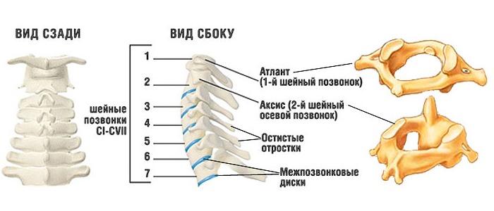
The human spine is made up of 24 vertebrae and four sections. . Each of them has significant differences in its structure and number of vertebrae. In the thoracic region, they are largest in size.
In the lumbar region, they are located very close to each other, and as they approach the coccygeal zone, they become fused. The cervical spine is considered the most fragile, but it is its thin structure that ensures the quality of mobility and allows you to make a variety of head movements.
The cervical region consists of seven vertebrae . Each of them differs in its structure. Due to their small size and weakness of the neck muscles, this section is often injured.
The peculiarity of the structure of the cervical vertebrae is significant differences from the vertebrae of all other parts of the spine. Most vertebrae consist of an anterior section called the vertebral body, which is cylindrical in shape; the spinal cord, located inside the spine from behind, is limited by the arch of the vertebra; they also have spinous processes pierced by holes for blood vessels.
The structure of the cervical vertebrae is different, which is due to the peculiarities of their functions, including fastening with the skull, protecting the spinal cord, providing nutrition to the brain and performing various head movements.
The structure and functions of the cervical vertebrae
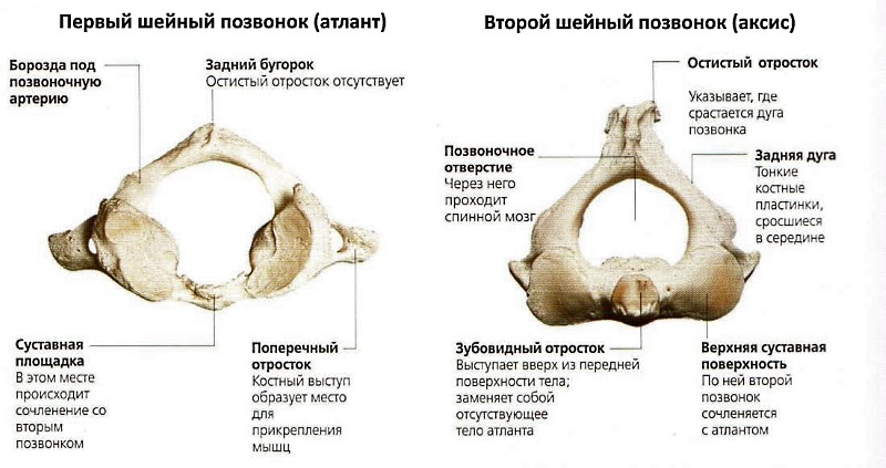
The very first vertebra of this department, located at the top, is called the atlas . It is axial, has no body and spinous process. In this area, it allows you to connect the spinal column with the bone of the neck, as well as the brain and spinal cord between themselves.
These tasks its structure is determined : it consists of two arches that border the spinal canal. The anterior arch forms a small tubercle. Behind it is a depression, combined with the odontoid process of the second vertebra.
A groove is located on the posterior arch, where the vertebral artery is placed.The articular part of the Atlanta, located on top, has a convex shape, and the lower – flat. Such a feature of the structure is due to the intermediate position of the vertebra between the spine and the head.
The second vertebra, called axis , also differs in its form, which resembles a pointed “tooth”. It performs the functions of a “hinge”, which provides the rotation of the first “Atlanta” vertebrae along with the skull, as well as the ability to perform the head tilments in different directions.
In the space between Atlant and Axis, there is no intervertebral disk . Their connection is formed by the type of joint. It is this factor that causes a high risk of injury.
Cervical vertebrae from the third to the sixth are small in size . Each of them has a fairly large hole, similar in shape to a triangle. Their upper edges are slightly protruding, due to which they are compared with the sides. Their articular processes are short and located at a small angle.
The vertebrae from the third to the fifth also have small transverse processes that are split at the edges. There are holes in these processes through which blood vessels pass. It is here that the main vertebral artery that feeds the brain is located.
On the next site, where the sixth and seventh vertebrae are located, the spinal column has a slight expansion . Here, the deposition of salts most often occurs. The sixth vertebra is called sleepy for the fact that its tubercle, located in front, is located near the cellular artery. It is to him that they press the artery to stop bleeding.
The largest in the last section of the cervical department is the seventh vertebra . It is it that you can feel it with your hands if you tilt your head forward. For the same reason, it is also called the speaker. In addition, it serves as the main guideline when calculating the vertebrae. The lower part of this vertebra has a recess.
Here is the place of its connection with the first rib. The peculiarity of the seventh vertebra is the holes in the field of transverse processes, which can be very small in size, or completely absent. It has the longest spicy growth, without divisions into parts.
Each of the vertebrae of the cervical region is responsible for a certain function.
With their damage, unpleasant phenomena arise that correspond to each specific vertebra, such as :
- Inflammatory and stagnant phenomena in the subordinate sinuses of the nose
- Soreness in the eyes
- Hearing deterioration
- Pain in the ears
- Facial nerve neuralgia
- whistles in the ears
- Face acne
- toothache
- caries
- bleeding gums
- Pain in the throat
- Chronic pharyngitis
- wheezing
- thyroid pathologies
- depression
- fears
- Pain in the shoulders
Permanent muscles of the cervical spine
Do you know that …
The muscle tissues of this spine are divided into two parts: the posterior and front.The muscles located in front are divided into superficial, deep and middle.
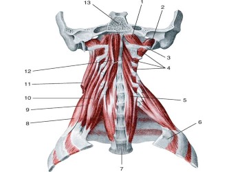 The main functions of tissues of the neck muscles are as follows :
- Support for the skull in equilibrium;
- ensuring the movement of the head: rotations and inclinations;
- Providing swallowing processes and voice function.
The muscle tissues of the cervical region are binded using special fascia and blood vessels, which serve as natural limiters of different areas.
Several main muscle groups are distinguished :
- subcutaneous muscles;
- muscles covering the surface of the neck;
- The shovel-key muscles necessary for the formation of the place, to place muscle tissues above the chest.
The muscles located inside the neck consist of the visceral plates necessary for the lining of organs inside the neck. They form areas in which veins and sleepy artery are located. The plate placed in front of the vertebra is needed to form a site to accommodate deep muscles.
Physiological bends of the cervical spine
The cervical spine has a natural bend directed forward . It is called lordosis. Such a bend is compensated by kyphosis – another bend directed back in the region of the thoracic region. Such bends give the spine elasticity, allow you to transfer daily loads due to straightforwardness.
The bends of the spine are not congenital. In order for them to correctly form the appropriate care and lifestyle.
Lordosis of the cervical department is considered physiological up to 40 degrees . If the angle exceeds this indicator, they diagnose
Anxiety should be taken and diagnosed in cases where the following symptoms of the development of pathology in the cervical spine were seen:
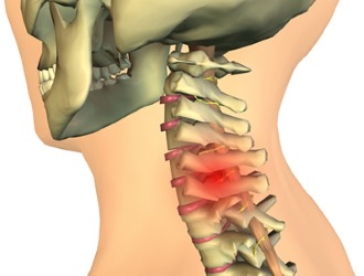
Diseases of the cervical region are possible due to various injuries after a severe blow or due to fall. In some cases, the danger of injury exists even with a sharp inclination or turning of the head, for example, when diving into water.
Most often, the following pathologies are found in the area of the cervical spine :
- ruptures of ligaments and intervertebral discs;
- vertebrae of vertebrae;
- Fractures.
Serious injuries of this department are dangerous in that they can hurt the spinal canal. The investigation may be paralysis, heart function or death. The danger of such injuries is also due to the fact that the whole seriousness of the situation can not always be evaluated immediately. At first, only pain during movement or swelling can indicate pathology.
Conclusion
The cervical spine It includes seven vertebrae, the structure of which differs markedly from the structure of the remaining sections of the spinal column.
Each of the vertebrae of this department performs certain functions. Damage to any of them can cause certain pathologies of the body.
The difference between these vertebrae lies in their small size and special fragility . Their shape is cylindrical, inside is the spinal cord.
The main functions of the cervical region are providing anchorage with the skull , nutrition for the brain, making a variety of head movements.
To ensure the same processes, the muscles of the neck serve, which also affect the processes of voice formation and swallowing.
The cervical region has a natural curve – lordosis , the correct formation of which occurs in the first years of human life and depends on the environment.
The most common diseases of the cervical spine are associated with various injuries, which are dangerous because they may not be noticed immediately, but they pose a risk of developing cardiac pathologies, paralysis, or even death.
Head and Neck Anatomy Exam Answers
(Faculty of Dentistry, 1st year) :
1. Anatomical norm. Anatomical variability: definition, classification, patterns of formation.
Anatomical norm – a genetically determined, rationally highly organized structure of the shape of the body, its organs, tissues and systems, which ensures the normal functioning of a person.
Development option – a deviation in the structure of the body within the normal range.
Anatomical variability (principles) :
ü determined by the influence of heredity and environment;
ü Implementation mechanism – processes of growth and development;
ü covers several signs at once;
associated with physiological variability;
ü leads to ambiguous results – variants of the structure of organs and the body.
MB: quantitative (change in size, mass) and qualitative.
Types of anatomical variability:
s age variability. Transformations in the process of ontogenesis – a change in the proportions of body parts, changes in the ratio of bones and muscles, changes in organs, etc.
Acceleration – acceleration of growth and development of the organism in the postnatal period of ontogenesis.
Change in diet, sedentary lifestyle, ecology, stress;
Retardation – slowdown in the growth and development of the organism in the postnatal period of ontogenesis.